

"Man is least himself when he talks in his own person. Give him a mask, and he will tell you the truth." Oscar Wilde
Showing posts with label CT scans. Show all posts
Showing posts with label CT scans. Show all posts
Tuesday, April 2, 2019
Diagnosing appendicitis


Thursday, November 1, 2018
Appendectomy outcomes in the modern era
Finally we have some data on the current rate of complications of appendectomy for uncomplicated appendicitis. But that’s not all. This new paper, published online in the journal Surgery, reveals much about the diagnosis, technique, and outcomes of appendectomy in the United States.
Using data from 115 hospitals participating in the National Surgical Quality Improvement Program, researchers at UCLA analyzed the results of 7778 adult patients undergoing appendectomy for simple appendicitis in 2016.
Using data from 115 hospitals participating in the National Surgical Quality Improvement Program, researchers at UCLA analyzed the results of 7778 adult patients undergoing appendectomy for simple appendicitis in 2016.
Thursday, August 23, 2018
A perforated colon case report raises a few issues
When a medical paper is featured on the Daily Mail website, you know it’s going to be something odd.
An autistic young man with prior hospitalizations for chronic constipation and megacolon was admitted to a hospital in London, UK with a markedly distended abdomen. A CT scan showed a dilated rectum and colon with a diameter as large as 18 cm (7 inches).

He was treated conservatively for two days with laxatives. Enemas were ordered, but the patient declined. He then developed peritonitis, kidney dysfunction, mental status changes, and metabolic acidosis.
An autistic young man with prior hospitalizations for chronic constipation and megacolon was admitted to a hospital in London, UK with a markedly distended abdomen. A CT scan showed a dilated rectum and colon with a diameter as large as 18 cm (7 inches).

He was treated conservatively for two days with laxatives. Enemas were ordered, but the patient declined. He then developed peritonitis, kidney dysfunction, mental status changes, and metabolic acidosis.
Monday, February 19, 2018
Perforated appendix not seen on CT scan
From an email received two days ago. Posted with permission and edited for length and clarity.
I came across your blog while looking for information on something puzzling that happened to my sister. I read the blog on CT scans and appendicitis* and went through all of the comments section. I couldn't find a case like my sister’s.
She has been sick off and on for the last two months. It began with what seemed like a bad stomach virus, fever, throwing up, diarrhea, gas pain, bloating, and stomach cramps. She brushed it off as a 24 hour type thing, stayed home from work a few days, and felt better but never returned to normal. She would have intermittent stomach upset and began to have weight loss. About 3 weeks ago, she went to see her GP who ran some tests (antibody tests, no CBC, chem 7 or normal work up) and diagnosed her with gluten intolerance.
She has been sick off and on for the last two months. It began with what seemed like a bad stomach virus, fever, throwing up, diarrhea, gas pain, bloating, and stomach cramps. She brushed it off as a 24 hour type thing, stayed home from work a few days, and felt better but never returned to normal. She would have intermittent stomach upset and began to have weight loss. About 3 weeks ago, she went to see her GP who ran some tests (antibody tests, no CBC, chem 7 or normal work up) and diagnosed her with gluten intolerance.
Monday, November 13, 2017
Useless general surgical interventions that should no longer be done
England’s National Health Service (NHS) could save €153 million [$178 million USD] per year by scrapping 71 low-value general surgery practices says a recently published paper in the British Journal of Surgery.
The authors, from Imperial College London, extensively reviewed the literature and Choosing Wisely recommendations from a number of countries.
I agree with many of the 71 including performing a cholecystectomy during the first admission for a patient with symptomatic gallstones instead of waiting for another admission (saving more than €54 million) and not repairing minimally symptomatic inguinal hernias, which they estimate would save over €32 million.
The Telegraph reported, “The team also discovered that robotic surgery has ‘little or no advantage’ when compared with traditional keyhole operations and said it must be ‘considered a candidate for disinvestment.’” This probably wouldn’t fly here in the US, because—like guns—too many robots are already in the hands of users.
The authors, from Imperial College London, extensively reviewed the literature and Choosing Wisely recommendations from a number of countries.
I agree with many of the 71 including performing a cholecystectomy during the first admission for a patient with symptomatic gallstones instead of waiting for another admission (saving more than €54 million) and not repairing minimally symptomatic inguinal hernias, which they estimate would save over €32 million.
The Telegraph reported, “The team also discovered that robotic surgery has ‘little or no advantage’ when compared with traditional keyhole operations and said it must be ‘considered a candidate for disinvestment.’” This probably wouldn’t fly here in the US, because—like guns—too many robots are already in the hands of users.
Wednesday, August 9, 2017
What to do when a normal looking appendix is found at surgery for appendicitis
For patients undergoing surgery with a presumptive diagnosis of appendicitis in Norway and other parts of Europe, the protocol is if the appendix looks grossly normal in the operating room, it is usually not removed.
This approach was mentioned as part of a paper on the readmission of post-appendectomy patients from Oslo University Hospital. Most of the patients underwent laparoscopy based on clinical diagnosis with only 160 having CT scans and 67 having ultrasounds.
Of the 710 patients in the Oslo series, 94% of the appendectomies were done laparoscopically, and 111 had a normal appearing appendix at laparoscopy. The appendix was not removed in 88. The other 23 patients had appendectomies for various reasons, and those appendices were normal at pathology.
The cumulative rate of operating for what turned out to be a normal appendix (88 + 23 cases) was 15.6%, which the authors attributed to “the low use of preoperative CT” due to concerns about radiation exposure. That over 100 patients had unnecessary general anesthesia and surgery was apparently not a concern.
This approach was mentioned as part of a paper on the readmission of post-appendectomy patients from Oslo University Hospital. Most of the patients underwent laparoscopy based on clinical diagnosis with only 160 having CT scans and 67 having ultrasounds.
Of the 710 patients in the Oslo series, 94% of the appendectomies were done laparoscopically, and 111 had a normal appearing appendix at laparoscopy. The appendix was not removed in 88. The other 23 patients had appendectomies for various reasons, and those appendices were normal at pathology.
The cumulative rate of operating for what turned out to be a normal appendix (88 + 23 cases) was 15.6%, which the authors attributed to “the low use of preoperative CT” due to concerns about radiation exposure. That over 100 patients had unnecessary general anesthesia and surgery was apparently not a concern.
Friday, June 24, 2016
Irrational fear of CT scans in appendicitis
By Skeptical Scalpel and Saurabh Jha*
Simple appendicitis cannot be distinguished from complicated appendicitis by clinical examination and laboratory findings say Finnish investigators. They looked at data from their randomized prospective trial of antibiotics vs. surgery for treatment of appendicitis and concluded that only CT scans could reliably differentiate the two entities.
The study involved adult patients from 18 to 60 years old; 368 of whom had uncomplicated acute appendicitis and 337 had complicated appendicitis—appendicolith, perforation, or abscess.
Duration of symptoms, C-reactive protein, white blood cell count, and temperature were significantly different between simple and complicated appendicitis patients. However substantial overlap of values meant they were not helpful in predicting the presence of complicated appendicitis.
Receiver operating curves for C-reactive protein and temperature areas under the curve do not exceed 0.77. Combining these parameters did not improve accuracy.
The paper concluded that CT scanning is essential in diagnosing acute appendicitis and identifying simple and complicated cases.
A companion study by many of the same authors looked at 1321 patients who presented with clinical and laboratory findings of possible appendicitis. Since their protocol called for confirmation of the diagnosis, all patients underwent CT scans, and 351 (27%) did not have appendicitis.
Simple appendicitis cannot be distinguished from complicated appendicitis by clinical examination and laboratory findings say Finnish investigators. They looked at data from their randomized prospective trial of antibiotics vs. surgery for treatment of appendicitis and concluded that only CT scans could reliably differentiate the two entities.
The study involved adult patients from 18 to 60 years old; 368 of whom had uncomplicated acute appendicitis and 337 had complicated appendicitis—appendicolith, perforation, or abscess.
Duration of symptoms, C-reactive protein, white blood cell count, and temperature were significantly different between simple and complicated appendicitis patients. However substantial overlap of values meant they were not helpful in predicting the presence of complicated appendicitis.
Receiver operating curves for C-reactive protein and temperature areas under the curve do not exceed 0.77. Combining these parameters did not improve accuracy.
The paper concluded that CT scanning is essential in diagnosing acute appendicitis and identifying simple and complicated cases.
A companion study by many of the same authors looked at 1321 patients who presented with clinical and laboratory findings of possible appendicitis. Since their protocol called for confirmation of the diagnosis, all patients underwent CT scans, and 351 (27%) did not have appendicitis.
Tuesday, September 16, 2014
Aortic dissection leads to man's death in the ED: His wife's perspective
A woman wrote to me about the day her husband died. I have edited her email for length and clarity and changed some insignificant details to protect her anonymity as she requested.
Joe passed away outside in the parking lot while they were getting on a helicopter for transport to a hospital equipped to do his surgery.
He had presented to the ED in terrible pain with lots of thrashing and writhing. His right hand was very cold. His right arm tingled to the point of hurting bad. The vision in his right eye was cloudy, and his hearing was muffled on the right. This was in addition to being very pale and diaphoretic upon admission. This is when I felt a dissecting aorta should have been suspected.
I don’t recall the vitals in the beginning, but they were changing and his blood pressure was dropping very fast. As soon as they finished the EKG-in the first 5 minutes of the visit, I asked the doctor about John Ritter's death [the actor died of a dissecting thoracic aneurysm in 2003]. First I asked if he could check for the condition that caused John Ritter's death. I called it an abdominal aortic aneurysm. The doc corrected me and said that it wasn’t an AAA it was a dissected aorta. I said OK, then check for that. This was 1 hour before the CT scan that led to his diagnosis.
Joe passed away outside in the parking lot while they were getting on a helicopter for transport to a hospital equipped to do his surgery.
He had presented to the ED in terrible pain with lots of thrashing and writhing. His right hand was very cold. His right arm tingled to the point of hurting bad. The vision in his right eye was cloudy, and his hearing was muffled on the right. This was in addition to being very pale and diaphoretic upon admission. This is when I felt a dissecting aorta should have been suspected.
I don’t recall the vitals in the beginning, but they were changing and his blood pressure was dropping very fast. As soon as they finished the EKG-in the first 5 minutes of the visit, I asked the doctor about John Ritter's death [the actor died of a dissecting thoracic aneurysm in 2003]. First I asked if he could check for the condition that caused John Ritter's death. I called it an abdominal aortic aneurysm. The doc corrected me and said that it wasn’t an AAA it was a dissected aorta. I said OK, then check for that. This was 1 hour before the CT scan that led to his diagnosis.
Wednesday, September 3, 2014
Health Care and the $20,000 Bruise: A different take
Twitter is buzzing about yet another medical billing horror story. This one appeared in the Wall Street Journal and was written by Eric Michael David who is an MD PhD JD and an officer at a biotech company.
He saw a large, swollen bruise on his three-year-old son's head several days after falling off his scooter. Other than the bruise, no other abnormalities were mentioned. He took the boy to "one of the top pediatric emergency rooms in the country" to have a CT scan done. It showed "a small, 11-day-old bleed inside his head, which was healing, and insignificant."
Dr. David received a bill for $20,000, $17,000 of which had been paid by his insurance company. He was responsible for the remaining $3000.
He noted a $10,000 charge for a trauma team activation which he said never happened. After a lengthy series of exchanges with the hospital's billing department and Dr. David having to prove that a trauma team activation was unwarranted and not permitted by certain regulations, he was able to have the charge rescinded.
The essay went on for some 1200 words listing the steps that he went through. He correctly described what a mess American healthcare delivery is and why as long as overuse and upcoding are rewarded, the Affordable Care Act will not fix it.
Dr. David was right to contest the $10,000 charge for a trauma team activation that wasn't indicated and didn't even occur.
What he didn't address was this.
Why would a doctor who said that he had "served on trauma teams in two of the busiest hospitals in New York City" feel the need to take his apparently asymptomatic son with an 11-day-old injury to an emergency room for a CT scan?
Doesn't this imply overuse of a different type?
Secondary questions:
Did anyone bring up the issue of radiation from the CT scan?
Did the docs in the ED think a CT scan was necessary?
"Inside his head" is a rather odd phrase. Does it mean intracranial? Intracerebral?
Was "one of the top pediatric emergency rooms in the country" the only option or could this asymptomatic boy have been seen in a doctor's office?
Why is the charge for a trauma team activation $10,000?
He saw a large, swollen bruise on his three-year-old son's head several days after falling off his scooter. Other than the bruise, no other abnormalities were mentioned. He took the boy to "one of the top pediatric emergency rooms in the country" to have a CT scan done. It showed "a small, 11-day-old bleed inside his head, which was healing, and insignificant."
Dr. David received a bill for $20,000, $17,000 of which had been paid by his insurance company. He was responsible for the remaining $3000.
He noted a $10,000 charge for a trauma team activation which he said never happened. After a lengthy series of exchanges with the hospital's billing department and Dr. David having to prove that a trauma team activation was unwarranted and not permitted by certain regulations, he was able to have the charge rescinded.
The essay went on for some 1200 words listing the steps that he went through. He correctly described what a mess American healthcare delivery is and why as long as overuse and upcoding are rewarded, the Affordable Care Act will not fix it.
Dr. David was right to contest the $10,000 charge for a trauma team activation that wasn't indicated and didn't even occur.
What he didn't address was this.
Why would a doctor who said that he had "served on trauma teams in two of the busiest hospitals in New York City" feel the need to take his apparently asymptomatic son with an 11-day-old injury to an emergency room for a CT scan?
Doesn't this imply overuse of a different type?
Secondary questions:
Did anyone bring up the issue of radiation from the CT scan?
Did the docs in the ED think a CT scan was necessary?
"Inside his head" is a rather odd phrase. Does it mean intracranial? Intracerebral?
Was "one of the top pediatric emergency rooms in the country" the only option or could this asymptomatic boy have been seen in a doctor's office?
Why is the charge for a trauma team activation $10,000?
Friday, June 13, 2014
Uncertain diagnosis or CT scan radiation? Which would you choose?
It is so nice to be right.
To summarize what I wrote almost 4 years ago, here
and here—based
on my experience, patients and families will accept the theoretical risk of a
future cancer if it means they'll get an accurate diagnosis.
A recent study
validates that opinion.
MedPage
Today reports that before receiving any recommendation for CT scanning, 742 parents of children who presented with head injuries
were surveyed by researchers from Toronto's Hospital for Sick Children.
Parents, almost half of whom had previously known that CT scanning
might cause a cancer to develop in the future, were told of the radiation risks
of CT scanning in detail. The authors found that although the parents'
willingness to go ahead with the CT scan fell from 90% before the explanation
of risk to 70% after they were briefed about radiation, at crunch time only 42
(6%) of them refused to let their child be scanned.
And of the 42 who initially refused, 8 eventually went ahead
with the scan after a physician recommended it.
So to put it another way, even after they were fully
informed of the potential risk of CT scan radiation to their child (lifetime
risk of cancer is about 1 in 10,000, according to the authors), nearly all parents
opted for the scan.
Also of note are the following:
The median age of the children was 4.
12% of the children in the study had undergone at least one
previous CT scan.
97% of the children were diagnosed with only concussions or
mild head injuries.
An article
in Scientific American puts some of the radiation risk into perspective. It is
long, but worth reading as it explains how risk has been calculated, the best
guess as to the true level of risk, and what radiologists are doing to lower
the radiation exposure associated with CT scanning.
According to that article, "Any one person in the U.S.
has a 20 percent chance of dying from cancer [of any type]. Therefore, a single
CT scan increases the average patient's risk of developing a fatal tumor from
20 to 20.05 percent."
No one ever comments about weighing the potential harms that may have been prevented by a timely CT scan diagnosis against the radiation risk.
CT scans should be ordered judiciously. The
area scanned and the amount of radiation should be limited as much as possible.
But if you need a CT scan to help diagnose your problem, go
ahead and have it.
Bottom line. When it comes to accuracy in diagnosis vs. radiation-induced
cancer risk, parents overwhelmingly chose the former.
Tuesday, June 25, 2013
What was surgery like in the 1970s?
When I first started my residency in the early 1970s, things were remarkably primitive by today's standards.
There were no ultrasound machines. Believe it or not, we would diagnose acute cholecystitis by history and physical examination alone. The only diagnostic tests we had were oral cholecystogram (OCG) and intravenous cholangiogram (IVC). For OCG, pills were taken the night before the test. If the cystic duct was patent, iodinated contrast would appear in the gallbladder and stones could be seen. Non-visualization of the gallbladder meant either the cystic duct was blocked or the pills were not absorbed (presumably due to inflammation, not necessarily of the GB) or the patient forgot to take the pills. The test was useless in acutely presenting patients. IVC was similar except the contrast was given intravenously. The common bile duct could be seen faintly unless the patient was jaundiced. It rarely showed stones in the GB.
There were no CT scans. We had to make the diagnosis of appendicitis by, you guessed it, history and physical examination alone. And since laparoscopic general surgery did not become common in the US until 1990, all appendectomies and cholecystectomies were done as open procedures.
There were no computers in any clinical departments or nursing units. Everything was on paper. The good news? There was no way to "copy and paste" progress notes. We had different colored paper for different sections of the chart, which made things easy to find. The bad news? Charts often went missing. Handwriting analysis rivaled that of archeologists deciphering hieroglyphics in Egypt. But paper charting was faster to do and easier to "leaf" through.
When submitting a research paper, drafts had to be prepared on a typewriter (an ancient kind of word processor that put the words directly on paper). If you needed to change a paragraph on page 1, the entire manuscript had to be retyped from the beginning. And making slides for presentations involved cameras with 35 mm film, taking the film to be developed and hoping the slides came out OK. Find a typo? Take the picture over and have the film processed again.
Now we use PowerPoint. It's easier, but I'm not totally convinced that it's real progress.
Maybe the biggest change has been the advent of the Internet. In the palm of my hand, I can instantly access huge amounts of information formerly available only in print books and journals. To look up a paper, we had to use Index Medicus, an encyclopedic series of books listing every article by subject in most journals.
There were far fewer journals back then. You had to know the correct heading or keyword to search or you could miss something important. Cross-referencing was not easy because it was in print and there was a different set of volumes for every year. And libraries kept many years' worth of volumes of journals.
Of course, many more changes have occurred. Can any of my older colleagues comment?
There were no ultrasound machines. Believe it or not, we would diagnose acute cholecystitis by history and physical examination alone. The only diagnostic tests we had were oral cholecystogram (OCG) and intravenous cholangiogram (IVC). For OCG, pills were taken the night before the test. If the cystic duct was patent, iodinated contrast would appear in the gallbladder and stones could be seen. Non-visualization of the gallbladder meant either the cystic duct was blocked or the pills were not absorbed (presumably due to inflammation, not necessarily of the GB) or the patient forgot to take the pills. The test was useless in acutely presenting patients. IVC was similar except the contrast was given intravenously. The common bile duct could be seen faintly unless the patient was jaundiced. It rarely showed stones in the GB.
There were no CT scans. We had to make the diagnosis of appendicitis by, you guessed it, history and physical examination alone. And since laparoscopic general surgery did not become common in the US until 1990, all appendectomies and cholecystectomies were done as open procedures.
There were no computers in any clinical departments or nursing units. Everything was on paper. The good news? There was no way to "copy and paste" progress notes. We had different colored paper for different sections of the chart, which made things easy to find. The bad news? Charts often went missing. Handwriting analysis rivaled that of archeologists deciphering hieroglyphics in Egypt. But paper charting was faster to do and easier to "leaf" through.
When submitting a research paper, drafts had to be prepared on a typewriter (an ancient kind of word processor that put the words directly on paper). If you needed to change a paragraph on page 1, the entire manuscript had to be retyped from the beginning. And making slides for presentations involved cameras with 35 mm film, taking the film to be developed and hoping the slides came out OK. Find a typo? Take the picture over and have the film processed again.
Now we use PowerPoint. It's easier, but I'm not totally convinced that it's real progress.
Maybe the biggest change has been the advent of the Internet. In the palm of my hand, I can instantly access huge amounts of information formerly available only in print books and journals. To look up a paper, we had to use Index Medicus, an encyclopedic series of books listing every article by subject in most journals.
There were far fewer journals back then. You had to know the correct heading or keyword to search or you could miss something important. Cross-referencing was not easy because it was in print and there was a different set of volumes for every year. And libraries kept many years' worth of volumes of journals.
Of course, many more changes have occurred. Can any of my older colleagues comment?
Wednesday, October 31, 2012
Can you guess the diagnosis using this radiology report?
Here are some excerpts from a radiology report I received. I
left out some portions that were not pertinent and I highlighted in bold some of the
better parts. My comments are in brackets.
This was in reference to a CT scan of the abdomen for
abdominal pain.
Free peritoneal air is present. Proximal small bowel loops are dilated to about 4 cm in diameter. In
the lower abdomen and upper pelvis there is a region of edema and a change in
caliber of the diameter of the bowel consistent
with bowel obstruction. This edema also surrounds the sigmoid colon. There
are diverticula and diverticulitis is a
consideration. Perforated
diverticulitis is also another consideration for the free air. There are
some collections of air along the right side of the sigmoid colon which may be extraluminal or perhaps some prominent diverticula. A
small abscess in this region is a consideration.
There is diffuse edema in the lower omentum.
No abnormal masses are
seen in the pelvis. No significant amount of free pelvic fluid is present.
{Is there an insignificant amount of fluid?]
No appendicitis. No diverticulitis. No free air.[!]
IMPRESSION: Proximal
small bowel obstruction. Questionable
diverticulitis with questionable small
abscess adjacent to the sigmoid colon. Pneumoperitoneum
[“Pneumoperitoneum” means free air in the abdominal cavity.]
Is there diverticulitis? Is there free air? What the hell is
going on?
So if you were the surgeon, what would you do?
At least the radiologist used the correct plural form of the
word “diverticulum.” See my previous post
on this topic.
Friday, October 12, 2012
Appendicitis: Accurate diagnosis or no radiation. You make the call.
Amid the mounting concern about radiation exposure and future increases in cancer rates comes a report from Washington State describing the benefits of imaging, particularly CT scanning, for the diagnosis of appendicitis.
The authors collected data from some 55 hospitals of all types and sizes over a six-year period for more than 19,300 patients older than age 15; 91% of patients underwent one or more imaging studies. There were 16,852 (87.2%) CT scans, 1160 (6.0%) ultrasounds and 108 MRIs (0.6%) with 1677 (8.7%) patients having no imaging.
If a patient had an imaging study, the rate of normal appendix removal was only 4.5% compared to 15.4% for those who were not imaged, p < 0.001. Stated another way, on multivariate analysis a patient who had no imaging was over 3 times more likely to have a normal appendix at surgery. Only 4.1% of those undergoing CT scan had a normal appendix compared to 10.4% of those who had only an ultrasound, p < 0.001.
The difference in the rate of normal appendix removal was true regardless of gender—men 3% with imaging vs. 10% without and women 6.9% vs. 24.7%, both statistically significant differences. In other words, nearly 1/4 of all women who were not imaged had a normal appendix at surgery.
Perforated appendicitis occurred in 15% of all patients, a figure that has remained constant over many years and has not been influenced by imaging.
The Washington appendicitis experience is probably generalizable because in most hospitals, CT scan is the preferred imaging modality over ultrasound, which is more operator-dependent, less accurate and less available during off hours than CT scan.
My personal experience is similar to that of the authors of the study. My rate of removal of a normal appendix when operating for the presumed diagnosis of appendicitis is 4.3% for my last 200 cases. Only 18 (9%) of my patients, all young males, had no imaging and just 1 had a normal appendix. CT scans were the only studies performed in 99% of my patients who were imaged.
I have previously written that patients and their families seem to prefer an accurate diagnosis over a theoretical slightly higher cancer risk 20 or 30 years later. The paper from Washington also confirms that a high accuracy rate of CT scanning for appendicitis is achievable outside of academic centers.
For many reasons, I can’t imagine the rate of CT scans for the diagnosis of patients with right lower quadrant pain decreasing. The accuracy of CT is no longer an issue. Most ED MDs are scanning everyone with right lower abdominal pain. They may tell you it’s because surgeons insist upon it, but whatever the reason, a lot of CT scans are being done. Another advantage of CT scanning is if the diagnosis is something other than appendicitis, the CT will very often reveal what is wrong with the patient. In a previous blog, I have mentioned before that it would be impractical to ask surgeons to examine every patient with abdominal pain, one of the more frequent complaints of patients presenting to emergency departments.
Face it, CT scanning for the diagnosis of appendicitis is here to stay. Whether implementation of protocols to lower the radiation dose of the devices will calm all the doomsday prophets remains to be seen.
[Thanks to Dr. Frederick Thurston Drake, lead author of the paper from Washington, for providing me with information not published.]
The authors collected data from some 55 hospitals of all types and sizes over a six-year period for more than 19,300 patients older than age 15; 91% of patients underwent one or more imaging studies. There were 16,852 (87.2%) CT scans, 1160 (6.0%) ultrasounds and 108 MRIs (0.6%) with 1677 (8.7%) patients having no imaging.
If a patient had an imaging study, the rate of normal appendix removal was only 4.5% compared to 15.4% for those who were not imaged, p < 0.001. Stated another way, on multivariate analysis a patient who had no imaging was over 3 times more likely to have a normal appendix at surgery. Only 4.1% of those undergoing CT scan had a normal appendix compared to 10.4% of those who had only an ultrasound, p < 0.001.
The difference in the rate of normal appendix removal was true regardless of gender—men 3% with imaging vs. 10% without and women 6.9% vs. 24.7%, both statistically significant differences. In other words, nearly 1/4 of all women who were not imaged had a normal appendix at surgery.
Perforated appendicitis occurred in 15% of all patients, a figure that has remained constant over many years and has not been influenced by imaging.
The Washington appendicitis experience is probably generalizable because in most hospitals, CT scan is the preferred imaging modality over ultrasound, which is more operator-dependent, less accurate and less available during off hours than CT scan.
My personal experience is similar to that of the authors of the study. My rate of removal of a normal appendix when operating for the presumed diagnosis of appendicitis is 4.3% for my last 200 cases. Only 18 (9%) of my patients, all young males, had no imaging and just 1 had a normal appendix. CT scans were the only studies performed in 99% of my patients who were imaged.
I have previously written that patients and their families seem to prefer an accurate diagnosis over a theoretical slightly higher cancer risk 20 or 30 years later. The paper from Washington also confirms that a high accuracy rate of CT scanning for appendicitis is achievable outside of academic centers.
For many reasons, I can’t imagine the rate of CT scans for the diagnosis of patients with right lower quadrant pain decreasing. The accuracy of CT is no longer an issue. Most ED MDs are scanning everyone with right lower abdominal pain. They may tell you it’s because surgeons insist upon it, but whatever the reason, a lot of CT scans are being done. Another advantage of CT scanning is if the diagnosis is something other than appendicitis, the CT will very often reveal what is wrong with the patient. In a previous blog, I have mentioned before that it would be impractical to ask surgeons to examine every patient with abdominal pain, one of the more frequent complaints of patients presenting to emergency departments.
Face it, CT scanning for the diagnosis of appendicitis is here to stay. Whether implementation of protocols to lower the radiation dose of the devices will calm all the doomsday prophets remains to be seen.
[Thanks to Dr. Frederick Thurston Drake, lead author of the paper from Washington, for providing me with information not published.]
Monday, April 18, 2011
Obesity: New Products and Old Problems
You might be interested in the response of equipment manufacturers to the increase in the size of obese patients. Below you can see a photo of a wide-body wheelchair and a super wide-body bedside commode.


You will note that the bedside commode has a normal sized bucket but the seat is extra wide to accommodate the girth of morbidly obese patients.
Something probably not appreciated by most people is that a morbidly obese person’s body actually contains a normal sized individual surround by a large layer of adipose tissue.
Below is an abdominal CT scan which illustrates the point.
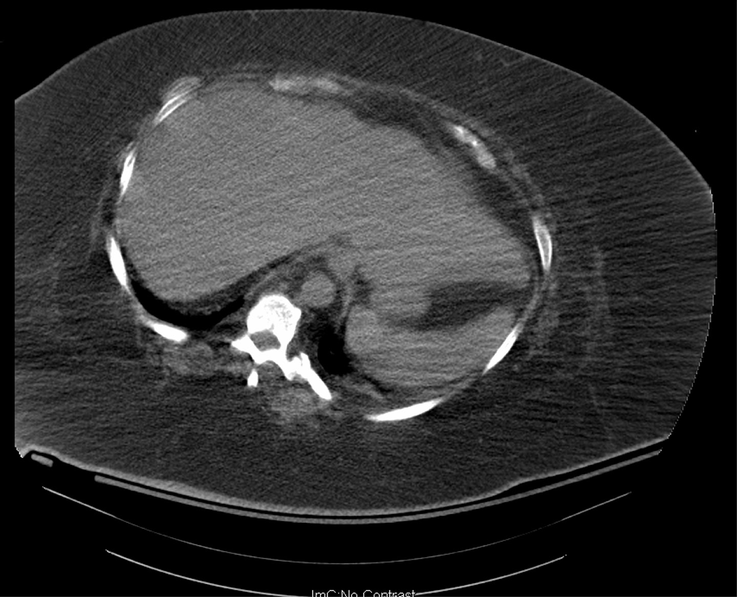
You can see that the organs are encased in body fat which extends to the limits (and beyond) of the CT scan table. The picture is grainy because the x-rays are partially absorbed by the thick fatty layer. This makes the interpretation of the images much more difficult. So not only is the diagnosis of abdominal problems hindered by the fat, you can imagine how hard surgery must be. Or maybe you can’t. Take my word for it, it’s hard.
Recovery is also an issue due to impaired wound healing, increased incidence of infections, problems moving the patient, difficult intravenous access and many more.
Remember this when you order that second Big Mac with supersized fries.
Monday, April 4, 2011
A Radiologist’s Idea of a Definitive Report
Here is an excerpt from the text of a CT scan report I received today:
The appendix is dilated and mildly thick walled with mild surrounding induration [sic]. There is gas within the lumen, but there is also an appendicolith at the base of the appendix with a smaller appendicolith in the mid appendix. The findings are concerning for appendicitis. No other inflammatory changes are seen. There is no adenopathy or free fluid.
IMPRESSION:
The appendix is dilated and mildly thick walled with suggestion of mild surrounding inflammatory change, although there is air within the lumen. There is an appendicolith at the base of the cecum. The findings are suspicious for mild acute appendicitis. Clinical and laboratory correlation are recommended.
I think it's somewhat mild. Don't you?
As the old joke goes, what is the radiologist’s favorite plant? The “hedge.”
The appendix is dilated and mildly thick walled with mild surrounding induration [sic]. There is gas within the lumen, but there is also an appendicolith at the base of the appendix with a smaller appendicolith in the mid appendix. The findings are concerning for appendicitis. No other inflammatory changes are seen. There is no adenopathy or free fluid.
IMPRESSION:
The appendix is dilated and mildly thick walled with suggestion of mild surrounding inflammatory change, although there is air within the lumen. There is an appendicolith at the base of the cecum. The findings are suspicious for mild acute appendicitis. Clinical and laboratory correlation are recommended.
I think it's somewhat mild. Don't you?
As the old joke goes, what is the radiologist’s favorite plant? The “hedge.”
Thursday, March 31, 2011
Too Many Diagnostic Tests? Says Who?
A recent Time magazine column attempted to analyze the reasons for the increased use of diagnostic imaging. The authors brought up many of the standard reasons including the need to practice defensive medicine, fallout from morbidity and mortality conferences, greed, etc. They say that doctors are no longer taught how to tell when patients need diagnostic tests and when they don’t. They make the following claim: “… the real value of so much testing has been widely questioned in scientific literature: imaging rates are going up, but doctors are not diagnosing (or necessarily misdiagnosing) more diseases.”
Respected medical blogger Kevin Pho (KevinMD) agreed with the Time article. Dr. Pho said that students are “…are spoiled by the easy access to the latest in imaging equipment and medical technology, and have become less dependent on physical exam skills previously relied upon in the past."
But as I have blogged previously, patients seem to want accurate diagnoses. Thus far, they are not moved by the theoretical increased risk of cancer secondary to radiation which may occur in the distant future. They want to know if they have appendicitis now and before they consent to surgery.
Contrary to what the Time magazine piece stated, there is some evidence that imaging does make the diagnosis of some diseases more accurate than history and physical examination.
In the April 2011 issue of the American Journal of Surgery, researchers from Howard University and the University of California, San Diego reported a 10-year review of negative appendectomy rates in the U.S. Using data from the National Inpatient Sample, they found that for nearly 500,000 appendectomies performed for presumed appendicitis from 1998 through 2007, the rate of removal of a normal appendix during surgery fell from 14.7% to 8.5%. This represents a 40% decline in the rate of negative appendectomy. The rate of perforated appendicitis also fell during that time period, indicating that the fall in negative appendectomy rates did not result in more occurrences of perforation.
Although the study was not designed to establish a link between the increased use of imaging studies and the falling rate of negative appendectomy, there are many papers supporting this conclusion such as one from Brigham and Women’s Hospital in Boston which showed a fall in the negative appendectomy rate from 23% to 1.7% over and 18-year period during which the rate of CT scan use went from 1% to 97.5%. Surgeons from Cornell reported similar results with the rate of CT scanning for appendicitis rising from 32% to 95% and the negative appendectomy rate falling from 16% to 7.65% from the late 1990s to the mid-part of the 21st century.
Having published a paper a few years ago which decried the use of CT scans in the diagnosis of appendicitis, I have completely reversed that opinion based on my personal series of over 125 appendectomies over the last two years with a negative appendectomy rate of just under 5%.
I never thought I would say this. The fact is CT scan is very accurate at diagnosing appendicitis. CT is better than I am and likely better than any human. It isn’t going to go away. It isn’t going to be replaced by ultrasound [not as readily available, not as accurate, not as easy to read] or MRI [not as readily available, not nearly as well-documented in the diagnosis of appendicitis, more expensive, not as easy to read]. So get over it.
Respected medical blogger Kevin Pho (KevinMD) agreed with the Time article. Dr. Pho said that students are “…are spoiled by the easy access to the latest in imaging equipment and medical technology, and have become less dependent on physical exam skills previously relied upon in the past."
But as I have blogged previously, patients seem to want accurate diagnoses. Thus far, they are not moved by the theoretical increased risk of cancer secondary to radiation which may occur in the distant future. They want to know if they have appendicitis now and before they consent to surgery.
Contrary to what the Time magazine piece stated, there is some evidence that imaging does make the diagnosis of some diseases more accurate than history and physical examination.
In the April 2011 issue of the American Journal of Surgery, researchers from Howard University and the University of California, San Diego reported a 10-year review of negative appendectomy rates in the U.S. Using data from the National Inpatient Sample, they found that for nearly 500,000 appendectomies performed for presumed appendicitis from 1998 through 2007, the rate of removal of a normal appendix during surgery fell from 14.7% to 8.5%. This represents a 40% decline in the rate of negative appendectomy. The rate of perforated appendicitis also fell during that time period, indicating that the fall in negative appendectomy rates did not result in more occurrences of perforation.
Although the study was not designed to establish a link between the increased use of imaging studies and the falling rate of negative appendectomy, there are many papers supporting this conclusion such as one from Brigham and Women’s Hospital in Boston which showed a fall in the negative appendectomy rate from 23% to 1.7% over and 18-year period during which the rate of CT scan use went from 1% to 97.5%. Surgeons from Cornell reported similar results with the rate of CT scanning for appendicitis rising from 32% to 95% and the negative appendectomy rate falling from 16% to 7.65% from the late 1990s to the mid-part of the 21st century.
Having published a paper a few years ago which decried the use of CT scans in the diagnosis of appendicitis, I have completely reversed that opinion based on my personal series of over 125 appendectomies over the last two years with a negative appendectomy rate of just under 5%.
I never thought I would say this. The fact is CT scan is very accurate at diagnosing appendicitis. CT is better than I am and likely better than any human. It isn’t going to go away. It isn’t going to be replaced by ultrasound [not as readily available, not as accurate, not as easy to read] or MRI [not as readily available, not nearly as well-documented in the diagnosis of appendicitis, more expensive, not as easy to read]. So get over it.
Monday, February 14, 2011
Statistics, Radiation and Childhood Cancers
A while ago, I tweeted that medical journalists should have a working knowledge of statistics. Perhaps that should be extended to those who perform peer review for journals as well. A report on Yahoo News via HealthDay [headlined “Greater Caution Urged for X-Rays in Pregnancy, Infants”] states “There's a small increased risk of cancer for children who had X-rays before they were 3 months old and those whose mothers had X-rays while pregnant, researchers say.”
A few paragraphs later comes this: “Children whose mothers had X-rays while pregnant had a slightly increased risk for all childhood cancers and for leukemia, though the increase was not statistically significant [emphasis added]. Children who had X-rays in early infancy had a small, non-significant [emphasis added] increased risk for all childhood cancers, leukemia and lymphoma.”
For those of you who still do not get it, an increased risk that is not statistically significant is the same as saying there is no difference in risk. A difference that is not statistically significant is simply not a difference at all. It could have happened by chance. It is misleading, if not downright dishonest, to state otherwise.
The paper actually does say that the risk for lymphoma only is statistically significantly increased but the odds ratio (odds ratio 5.14, 1.27 to 20.78) is quite wide due to the small numbers of patients in the study.
This was a case-control study of 2690 childhood cancer cases and 4858 age, sex, and region matched controls from the United Kingdom. It was published in the British Medical Journal on February 10, 2011.
There is heighten awareness in the literature about the risks of radiation from diagnostic tests. Real concern is warranted. But this report adds only noise and confusion.
A few paragraphs later comes this: “Children whose mothers had X-rays while pregnant had a slightly increased risk for all childhood cancers and for leukemia, though the increase was not statistically significant [emphasis added]. Children who had X-rays in early infancy had a small, non-significant [emphasis added] increased risk for all childhood cancers, leukemia and lymphoma.”
For those of you who still do not get it, an increased risk that is not statistically significant is the same as saying there is no difference in risk. A difference that is not statistically significant is simply not a difference at all. It could have happened by chance. It is misleading, if not downright dishonest, to state otherwise.
The paper actually does say that the risk for lymphoma only is statistically significantly increased but the odds ratio (odds ratio 5.14, 1.27 to 20.78) is quite wide due to the small numbers of patients in the study.
This was a case-control study of 2690 childhood cancer cases and 4858 age, sex, and region matched controls from the United Kingdom. It was published in the British Medical Journal on February 10, 2011.
There is heighten awareness in the literature about the risks of radiation from diagnostic tests. Real concern is warranted. But this report adds only noise and confusion.
Wednesday, February 2, 2011
Abdominopelvic CT Increases Diagnostic Certainty
A report from the Massachusetts General Hospital shows that abdominopelvic CT scans significantly increased diagnostic accuracy and changed patient management in a series of 584 prospectively studied non-trauma patients with abdominal pain. The paper was published ahead of print in the American Journal of Radiology.
Emergency physicians recorded their presumptive diagnoses after examining patients and before the CT scan was performed. Key findings were that CT scanning increased diagnostic certainty from 70% to 90% and resulted in changes in patient treatment plans in 42%. In addition, 25% of patients initially diagnosed with a problem thought to require surgery were discharged from the emergency department without further treatment.
The most common problems encountered were ureteral stones, bowel obstruction and no acute condition, which was thought to be the diagnosis in 77 patients before CT scanning. Post-CT scan, the number of patients with no acute condition was 174, representing a 126% increase in that finding.
The paper did not address cost or radiation exposure. Asked about radiation exposure, lead author Hani H. Abujudeh, M.D. said in an email, radiation to the patient can be decreased “…using the latest technologies [and] knowledge we have.”
A surgeon, who asked to remain anonymous, reviewed the paper and offered a different interpretation of the data. He said that based on the nearly 50% post-CT rate of change in the diagnoses of appendicitis and bowel obstruction, one could simply say that the emergency department physicians at MGH are not particularly astute diagnosticians. He went on to say that the use of CT scans for abdominal pain is so common at his hospital, “if you complain of abdominal pain to the [emergency department] triage nurse, you are given your [oral] contrast to drink while you are still in the waiting room!”
This paper will no doubt be cited as justification for the increasing use of CT scanning in the diagnosis of abdominal pain. The blogger Skeptical Scalpel has commented that the quest for diagnostic accuracy trumps concerns about radiation.
Emergency physicians recorded their presumptive diagnoses after examining patients and before the CT scan was performed. Key findings were that CT scanning increased diagnostic certainty from 70% to 90% and resulted in changes in patient treatment plans in 42%. In addition, 25% of patients initially diagnosed with a problem thought to require surgery were discharged from the emergency department without further treatment.
The most common problems encountered were ureteral stones, bowel obstruction and no acute condition, which was thought to be the diagnosis in 77 patients before CT scanning. Post-CT scan, the number of patients with no acute condition was 174, representing a 126% increase in that finding.
The paper did not address cost or radiation exposure. Asked about radiation exposure, lead author Hani H. Abujudeh, M.D. said in an email, radiation to the patient can be decreased “…using the latest technologies [and] knowledge we have.”
A surgeon, who asked to remain anonymous, reviewed the paper and offered a different interpretation of the data. He said that based on the nearly 50% post-CT rate of change in the diagnoses of appendicitis and bowel obstruction, one could simply say that the emergency department physicians at MGH are not particularly astute diagnosticians. He went on to say that the use of CT scans for abdominal pain is so common at his hospital, “if you complain of abdominal pain to the [emergency department] triage nurse, you are given your [oral] contrast to drink while you are still in the waiting room!”
This paper will no doubt be cited as justification for the increasing use of CT scanning in the diagnosis of abdominal pain. The blogger Skeptical Scalpel has commented that the quest for diagnostic accuracy trumps concerns about radiation.
Wednesday, November 10, 2010
Surprising common sense from website Cracked.com: “5 common medical procedures that secretly aren’t worth it”
Yesterday, the humor website Cracked.com published a list of 5 common medical procedures that, in its opinion, aren’t worth it. They are CT scans, physical examinations, circumcisions, Cesarean sections and antibiotics. OK, antibiotics are not really procedures, but you get the point. Of course, the author uses exaggeration to bolster his case but there is a lot of truth in the article. Let’s take them one by one.
5. CT scans. No doubt about it, they are overused and they are expensive. And there is a theoretical and possibly real risk, albeit small, of an increase in cancer rates in the future. Cracked thinks the increase in the use of CT scans is about money and greed but the fact is that doctors who order CT scans almost always do not own the scanner and do not profit from ordering the test. I think it’s about the public’s demand for diagnostic accuracy (I blogged about this in August) and the pervasive practice of defensive medicine. Physicians feel compelled to cover their asses by over-ordering confirmatory tests before doing anything and they fear being sued for missing something. As an alternative, Cracked.com suggests increasing the use of MRI. Although MRI does not involve radiation to the patient, it is also very expensive and not routinely available on a 24/7 basis. For some illnesses, it is just not as good as a CT scan for diagnosis.
4. Physical examinations. I could not agree more that they are useless in asymptomatic patients.
3. Circumcisions. More than 35 years ago, circumcision was shown to be not only unnecessary, but probably harmful in that occasionally a child was mutilated by technical errors with the procedure. Children with unsuspected hereditary bleeding disorders have even died during circumcision. Why the practice continues is inexplicable. I tweeted about this a few months ago and am proud to be now listed by the tweeter “Intact by Default” as disfavoring circumcision.
2. Cesarean sections. There are certainly too many Cesarean sections done in the U.S. Some are performed for the convenience of the patient or the obstetrician. However, the procedure can be “worth it” in selected patients. Examples of necessary Cesarean sections are the baby is too big to be delivered vaginally, the mother has a major complication of pregnancy or labor, some multiple births and others. Cesarean sections may also fall into the defensive medicine category as one can always question why a baby with a subsequent problem was delivered vaginally instead of by Cesarean section.
1. Antibiotics. Please, don’t get me started. Again although not a procedure, Cracked.com is spot on that antibiotics are vastly overused in medicine (and agriculture, by the way). In most hospitals, anyone who develops a fever gets antibiotics. Everyone wants antibiotics for a cold despite the fact that colds are caused by viruses (not affected by antibiotics) and colds are self-limited, non-fatal diseases. Overuse of antibiotics leads to bacterial resistance, which is now a major problem worldwide. They also cause diarrhea and a sometimes lethal infection called C. difficle colitis, which often attacks hospitalized, debilitated patients.
So let’s give Cracked.com an A- for its list. Too bad more physicians don’t read it. And too bad we live in a litigious society which fosters defensive medicine.
5. CT scans. No doubt about it, they are overused and they are expensive. And there is a theoretical and possibly real risk, albeit small, of an increase in cancer rates in the future. Cracked thinks the increase in the use of CT scans is about money and greed but the fact is that doctors who order CT scans almost always do not own the scanner and do not profit from ordering the test. I think it’s about the public’s demand for diagnostic accuracy (I blogged about this in August) and the pervasive practice of defensive medicine. Physicians feel compelled to cover their asses by over-ordering confirmatory tests before doing anything and they fear being sued for missing something. As an alternative, Cracked.com suggests increasing the use of MRI. Although MRI does not involve radiation to the patient, it is also very expensive and not routinely available on a 24/7 basis. For some illnesses, it is just not as good as a CT scan for diagnosis.
4. Physical examinations. I could not agree more that they are useless in asymptomatic patients.
3. Circumcisions. More than 35 years ago, circumcision was shown to be not only unnecessary, but probably harmful in that occasionally a child was mutilated by technical errors with the procedure. Children with unsuspected hereditary bleeding disorders have even died during circumcision. Why the practice continues is inexplicable. I tweeted about this a few months ago and am proud to be now listed by the tweeter “Intact by Default” as disfavoring circumcision.
2. Cesarean sections. There are certainly too many Cesarean sections done in the U.S. Some are performed for the convenience of the patient or the obstetrician. However, the procedure can be “worth it” in selected patients. Examples of necessary Cesarean sections are the baby is too big to be delivered vaginally, the mother has a major complication of pregnancy or labor, some multiple births and others. Cesarean sections may also fall into the defensive medicine category as one can always question why a baby with a subsequent problem was delivered vaginally instead of by Cesarean section.
1. Antibiotics. Please, don’t get me started. Again although not a procedure, Cracked.com is spot on that antibiotics are vastly overused in medicine (and agriculture, by the way). In most hospitals, anyone who develops a fever gets antibiotics. Everyone wants antibiotics for a cold despite the fact that colds are caused by viruses (not affected by antibiotics) and colds are self-limited, non-fatal diseases. Overuse of antibiotics leads to bacterial resistance, which is now a major problem worldwide. They also cause diarrhea and a sometimes lethal infection called C. difficle colitis, which often attacks hospitalized, debilitated patients.
So let’s give Cracked.com an A- for its list. Too bad more physicians don’t read it. And too bad we live in a litigious society which fosters defensive medicine.
Wednesday, September 29, 2010
Cancer Risk from CT Scan Exposure May Be Higher than You Think
According to two recent studies the cancer risk from radiation exposure may be higher than once thought. Berrington de Gonzalez and colleagues published two papers in the December 14/28, 2009 issue of Archives of Internal Medicine. One study postulates that CT scans in the U.S. in the year 2007 will lead to some 29,000 new cancers in the future. The largest number of cancers will result from CT scans of the abdomen.
The second paper investigated the variation in CT scan technique at four hospitals in the San Francisco area. The authors found wide variations in radiation dose due to different settings on the devices and the radiation exposure was much higher than previously published data suggested. For example, the median exposure from an abdominal CT scan was 31 millisieverts, a figure about four times higher than the usually quoted range.
For comparison, a CT scan of the abdomen and pelvis with contrast has an estimated radiation exposure to the patient of some 234 routine chest x-rays. A CT coronary angiogram exposes a patient to the same dose of radiation as 309 chest x-rays.
Editorialists in both Archives of Internal Medicine and JAMA expressed great concern and call for tighter regulation. The radiologists are also considering measures to try to decrease the number of CT scans done in the U.S.
The second paper investigated the variation in CT scan technique at four hospitals in the San Francisco area. The authors found wide variations in radiation dose due to different settings on the devices and the radiation exposure was much higher than previously published data suggested. For example, the median exposure from an abdominal CT scan was 31 millisieverts, a figure about four times higher than the usually quoted range.
For comparison, a CT scan of the abdomen and pelvis with contrast has an estimated radiation exposure to the patient of some 234 routine chest x-rays. A CT coronary angiogram exposes a patient to the same dose of radiation as 309 chest x-rays.
Editorialists in both Archives of Internal Medicine and JAMA expressed great concern and call for tighter regulation. The radiologists are also considering measures to try to decrease the number of CT scans done in the U.S.
Subscribe to:
Posts (Atom)